Acrodermatitis continua of Hallopeau in a 47-year-old female
SPMC J Health Care Serv. 2024;10(1):3 ARK: https://n2t.net/ark:/76951/jhcs7c5mq5
1Department of Dermatology, Southern Philippines Medical Center, JP Laurel Ave, Davao City, Philippines
Correspondence Kirk Llew V Quijote, kirkllewkirk@gmail.com
Received 18 January 2023
Accepted 6 June 2024
Cite as Quijote KLV, Segovia JCLQ, Castaños KPB, Visitacion LR. Acrodermatitis continua of Hallopeau in a 47-year-old female. SPMC J Health Care Serv. 2024;10(1):3. https://n2t.net/ark:/76951/jhcs7c5mq5
|
|
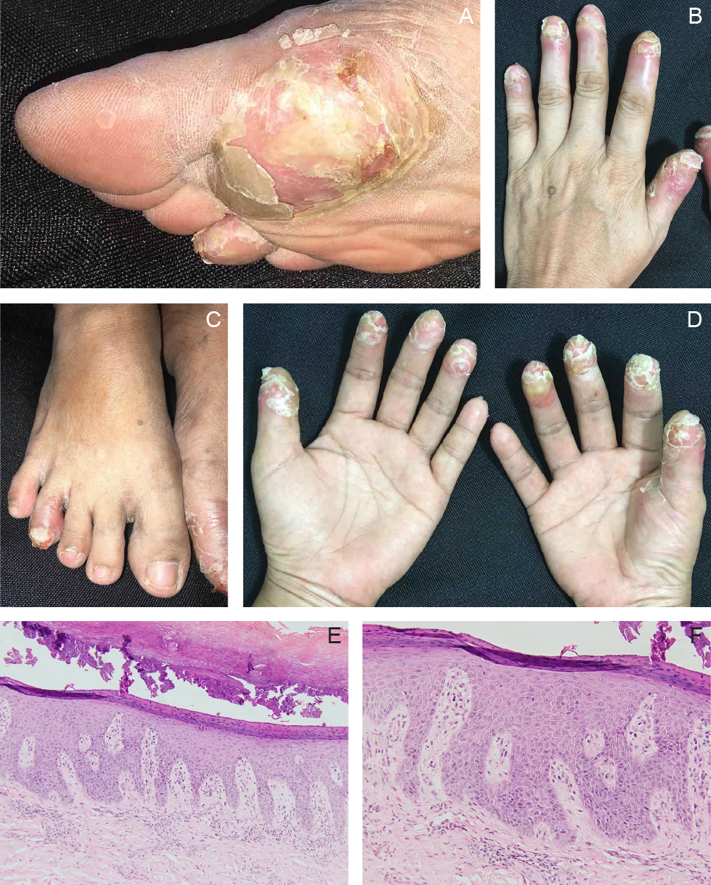
Figure 1 Clinical and histopathologic features of acrodermatitis continua of Hallopeau. Multiple erythematous pustules and thick white hyperkeratotic plaques with scales located on the distal interphalangeal joints extend to the tips of all digits of both hands. Similar lesions are also seen on the first metatarsophalangeal joint and the fourth digit of the right foot, and on the first digit of the left foot. Palmoplantar sparing and anonychia on all fingers and the left big toe are evident (A,B,C,D). Histopathology shows focal parakeratosis overlying a spongiotic dermis with hypogranulosis and psoriasiform hyperplasia (E: hematoxylin-eosin stain, x10). The dermis shows superficial dermal edema and moderately dense perivascular inflammatory infiltrates composed predominantly of lymphocytes and neutrophils (F: hematoxylin-eosin stain, x100). |
Contributors
KLVQ, JCLQS, KPBC, and LRV contributed to the diagnostic and therapeutic care of the patient in this report. All of them acquired relevant patient data, and searched for and reviewed relevant medical literature used in this report. KLVQ wrote the original draft, performed the subsequent revisions. All approved the final version, and agreed to be accountable for all aspects of this report.
Acknowledgments
We would like to thank Dr Cassandra Grace Bahjin, and Dr Brice Serquiña of the Department of Dermatology in Southern Philippines Medical Center for their help in the patient’s clinical follow-ups and management, as well as their contributions in providing resources for the write-up of the clinical images article.
Patient consent
Obtained
Reporting guideline used
Article source
Submitted
Peer review
External
Competing interests
None declared
Access and license
This is an Open Access article licensed under the Creative Commons Attribution-NonCommercial 4.0 International License, which allows others to share and adapt the work, provided that derivative works bear appropriate citation to this original work and are not used for commercial purposes. To view a copy of this license, visit https://creativecommons.org/licenses/by-nc/4.0/
References
1 Spiteri V, Bibra A, Ashwood N, Cobb J. Managing acrometastases treatment strategy with a case illustration. Ann R Coll Surg Engl. 2008 Oct;90(7):W8-11.
2 Balestri R, Magnano M, Ioris T, Girardelli CR, Rech G. Squamous cell carcinoma mimicking acrodermatitis continua suppurativa in a patient with generalized pustular psoriasis. J Dtsch Dermatol Ges. 2023 May;21(5):537-539.
3 Langley A, Asai Y. Pustules of the fingers: acrodermatitis continua. CMAJ. 2016 Oct 18;188(15):1105.
4 Kang S, Amagai M, Bruckner AL, Enk AH, Margolis DJ, McMichael AJ, et al, editors. Fitzpatrick's Dermatology, 9th ed. 2019. New York: McGraw Hill. Available from: https://accessmedicine.mhmedical.com/content.aspx?bookid=2570§ionid=210413306
5 Maliyar K, Crowley EL, Rodriguez-Bolanos F, O'Toole A, Gooderham MJ. The Use of Biologic Therapy in the Treatment of Acrodermatitis Continua of Hallopeau: A Review. J Cutan Med Surg. 2019 Jul/Aug;23(4):428-435.
6 Chularojanamontri L, Rattanakorn K, Julanon N, Chuamanochan M, Griffiths CEM. Acrodermatitis continua of Hallopeau and generalised pustular psoriasis: Should they be the same or different entities? Exp Dermatol. 2023 Aug;32(8):1235-1245.
7 Wen P, Liu C, Wang T, Jiang X, Wang P, Wang S. Successful treatment of acrodermatitis continua of Hallopeau coexisting with generalized pustular psoriasis with spesolimab: a case report. Front Immunol. 2024 Feb 23;15:1338285.
8 Chen YL, Wang ZY, Ma L, Xu ZG. Three cases of IL36RN-associated pustulosis: An evolution of acrodermatitis continua of Hallopeau to generalized pustular psoriasis. Indian J Dermatol Venereol Leprol. 2020 Sep-Oct;86(5):562-565.
9 Navarini AA, Burden AD, Capon F, Mrowietz U, Puig L, Köks S, et al. European consensus statement on phenotypes of pustular psoriasis. J Eur Acad Dermatol Venereol. 2017 Nov;31(11):1792-1799.
10 Mitra D, Bhatnagar A, Kumar M. Acrodermatitis Continua of Hallopeau: A Diagnostic Challenge. Indian Dermatol Online J. 2022 Dec 14;14(1):91-93.
11 Sehgal VN, Verma P, Sharma S, Srivastava G, Aggarwal AK, Rasool F, et al. Acrodermatitis continua of Hallopeau: evolution of treatment options. Int J Dermatol. 2011 Oct;50(10):1195-211.
12 Kromer C, Loewe E, Schaarschmidt ML, Pinter A, Gerdes S, Celis D, et al. Treatment of acrodermatitis continua of Hallopeau: A case series of 39 patients. J Dermatol. 2020 Sep;47(9):989-997.
13 Cirone KD, Lovegrove FE. Acrodermatitis continua of Hallopeau successfully treated with bimekizumab: A case report. SAGE Open Med Case Rep. 2023 Mar 21;11:2050313X231160937.


This work is licensed under a Creative Commons Attribution-NonCommercial 4.0 International License.
Authors who publish with this journal agree to the following terms:
- Authors retain copyright and grant the journal right of first publication with the work simultaneously licensed under a Creative Commons Attribution-NonCommercial 4.0 International License that allows others to share the work for non-commercial purposes with an acknowledgement of the work's authorship and initial publication in this journal.
- Authors are able to enter into separate, additional, non-commercial contractual arrangements for the non-exclusive distribution of the journal's published version of the work (e.g., post it to an institutional repository or publish it in a book), with an acknowledgement of its initial publication in this journal.
- Authors grant the journal permission to rewrite, edit, modify, store and/or publish the submission in any medium or format a version or abstract forming part thereof, all associated supplemental materials, and subsequent errata, if necessary, in a publicly available publication or database.
- Authors warrant that the submission is original with the authors and does not infringe or transfer any copyright or violate any other right of any third parties.